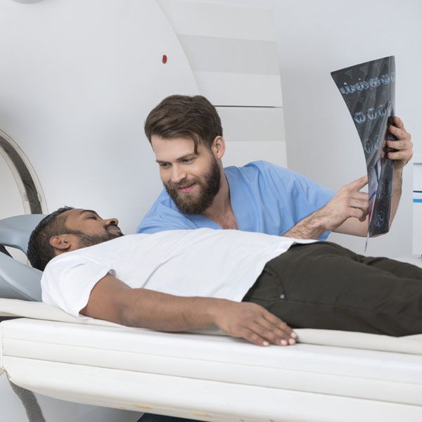
Abdominal Interventions
Interventional Radiologists at Triad Radiology Associates use imaging such as xray, CT, MRI, and ultrasound to provide a full range of diagnostic and therapeutic treatment options related to the chest, abdomen, pelvis, gastrointestinal, and genitourinary tracts.
Transjugular Intrahepatic Portosystemic Shunt (TIPS) Creation
Interventional Radiologists use image guidance to make a tunnel through the liver to connect the portal vein (the vein that carries blood from the digestive organs to the liver) to one of the hepatic veins (the vein that carry blood away from the liver back to the heart). A stent is then placed in this tunnel to keep the pathway open.
A TIPS procedure is used to treat complications of portal hypertension (increased
pressure in the portal vein system) including:
• Variceal bleeding — bleeding from any of the veins that normally drain the stomach, esophagus, or intestines into the liver
• Severe ascites and/or hydrothorax — accumulation of fluid in the abdomen and/or chest
• Portal gastropathy — an engorgement of the veins that line the stomach, which can result in severe bleeding
• Budd-Chiari syndrome — a blockage in one or more veins that carry blood from the liver back to the heart
Percutaneous Cholecystostomy
Cholecystitis is the inflammation of the gallbladder (the small organ under your liver), which typically occurs when the passage that connects the gallbladder to the bile duct is blocked. It may be acute (sudden inflammation that causes severe pain in the upper abdomen) or chronic (swelling and irritation that persists over time).
In most circumstances, surgical removal of the gallbladder is recommended for resolution of symptoms. In patients who are too ill to undergo surgical removal of the gallbladder, Interventional Radiologists use minimally invasive image guided techniques to place a small tube directly through the skin into the gallbladder in order to allow the flow of congested or infected bile to the outside, so that inflammation of the gallbladder can be reduced.
Percutaneous Transhepatic Cholangiogram (PTC) with Drainage
Percutaneous transhepatic cholangiography (PTC), a type of biliary intervention, is an xray procedure that involves the injection of a contrast material directly into the bile ducts inside the liver to produce pictures of the bile ducts. Biliary interventions are minimally invasive procedures performed to treat blockages, narrowing, and injury of bile ducts.
If a blockage or narrowing is found, additional procedures may be performed, including:
• insertion of a catheter to drain excess bile out of the body.
• removal of gallstones and debris that form in the gallbladder or bile ducts.
• stent placement inside a duct to help it remain open or to bypass an obstruction and allow fluids to drain internally.
Percutaneous Nephrostomy and Ureteral Stent Placement
The kidneys drain into the ureters, which are long narrow tubes that carry urine to the bladder. Ureters can become obstructed due to conditions such as kidney stones, tumors, infection, or blood clots. When this happens, Interventional Radiologists can use image guidance to place stents or thin flexible tubes into the ureter to restore urine flow.
When it is not possible to insert an ureteral stent, nephrostomy is performed. During this procedure, a tube is placed through the skin on the patient’s back into the kidney. The tube is connected to an external drainage bag, which helps to divert urine flow and to preserve renal function.
Suprapubic Catheter Placement
A suprapubic catheter (SPC) drains urine from your bladder.You may need a catheter because you have urinary incontinence (leakage), urinary retention (not being able to urinate), surgery that made a catheter necessary, or another health problem.
Normally, a catheter is inserted into your bladder through your urethra. A SPC, however, is inserted into your bladder through a small hole in your belly, just above your pubic bone. This allows urine to be drained or diverted outside to an external bag without having a tube in your genital area.
Your physician will instruct you on how to care for your catheter and how to ensure that it is working properly.
G and GJ Feeding Tube Insertion
Gastrostomy (G) or gastrojejunostomy (GJ) feeding tube insertion is the placement of a feeding tube to help with malnutrition or difficulty swallowing which can happen with cancer or strokes.
Gastrostomy tube is a feeding tube placed through the skin of the abdomen into the stomach. Gastrojejunostomy tube, on the other hand, is placed through the abdominal wall into the stomach and then through the duodenum into the jejunum.
Sources:
-
- https://www.radiologyinfo.org/en/info.cfm?pg=tips
- https://www.radiologyinfo.org/en/info.cfm?pg=cholecystitis
- https://www.radiologyinfo.org/en/info.cfm?pg=biliary
- https://www.radiologyinfo.org/en/info.cfm?pg=ureteralNephro
- https://medlineplus.gov/ency/patientinstructions/000145.htm
- http://www.childrenshospital.org/conditions-and-treatments/treatments/primary-percutaneous-gastrojejunostomy
Triad Radiology offers Abdominal Interventions at a variety of locations, including hospitals, imaging centers, and clinics. Contact us if you want to learn more or schedule an appointment.


